
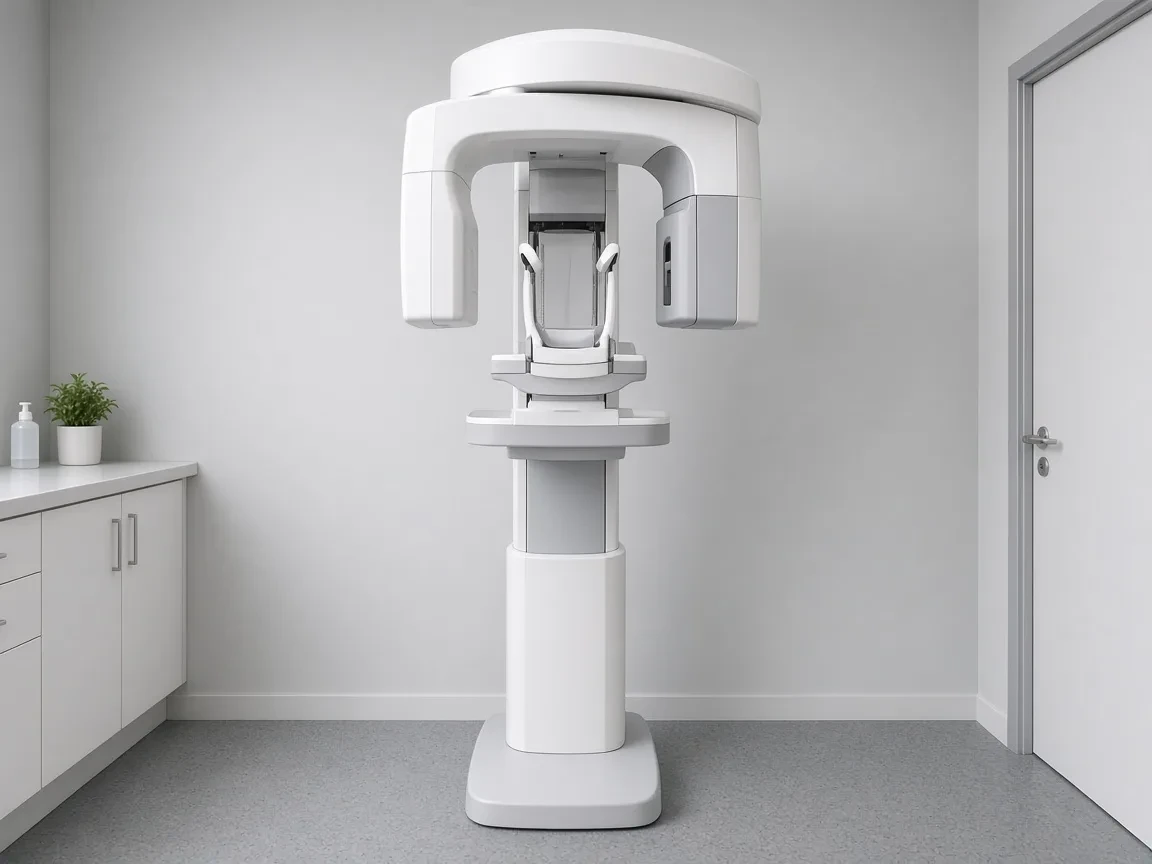
Imaging
Dental X-ray units, sensors, phosphor plate systems, panoramic units, CBCT and intraoral cameras, compared for the exams a general practice takes most.
Summary
These guides cover the equipment a practice uses to take and view dental images: intraoral X-ray units, digital sensors, phosphor plate systems, panoramic units, CBCT and intraoral cameras. Each one compares the main brands and explains which exams the device suits, so you can match the purchase to the images your patients need.
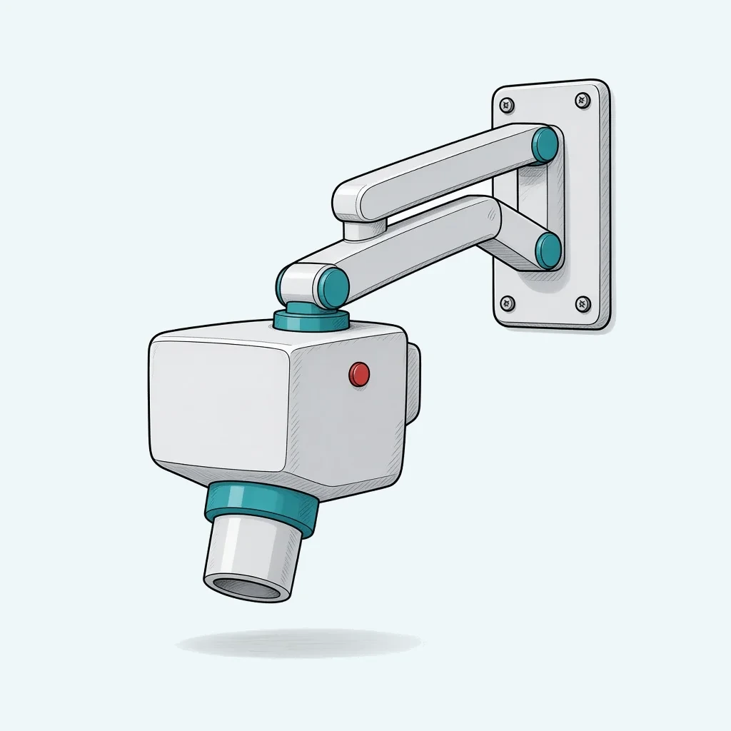
Intraoral X-ray Units: Wall-Mount vs Handheld NOMAD
A wall-arm or handheld intraoral x-ray unit, a DC generator, and how Veraview, ProX, and NOMAD differ.
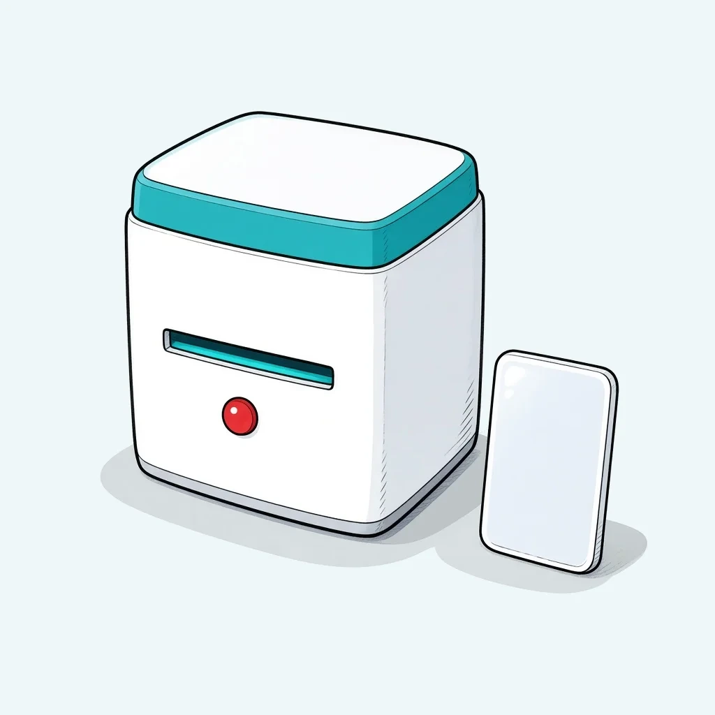
Phosphor Plate Systems: ScanX, VistaScan, Xios Scan
Phosphor plate systems: reusable plates, a reader that is not the x-ray head, and how ScanX, Xios, and Pspix differ.
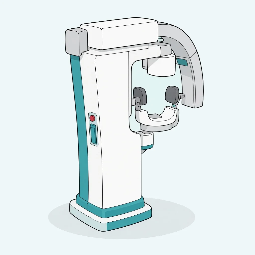
Dental CBCT Systems: Field of View, Dose and Brands
CBCT field of view, voxel size, and 2D modes, and how Planmeca, Dentsply Sirona, Carestream, Vatech, DEXIS, and Morita differ.
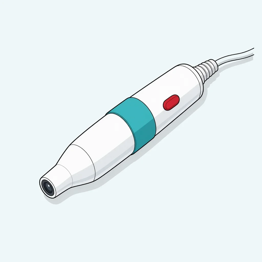
Intraoral Cameras: MouthWatch, DEXIS, Acteon Compared
Intraoral cameras compared on focus, capture, and software: MouthWatch, DEXIS, Acteon, Digital Doc, and Carestream.
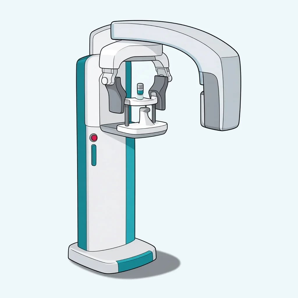
Panoramic X-ray Units: Planmeca, Vatech, Carestream
2D panoramic versus combo CBCT, ceph, and patient positioning, and how Planmeca, Dentsply Sirona, Vatech, Carestream, and DEXIS differ.
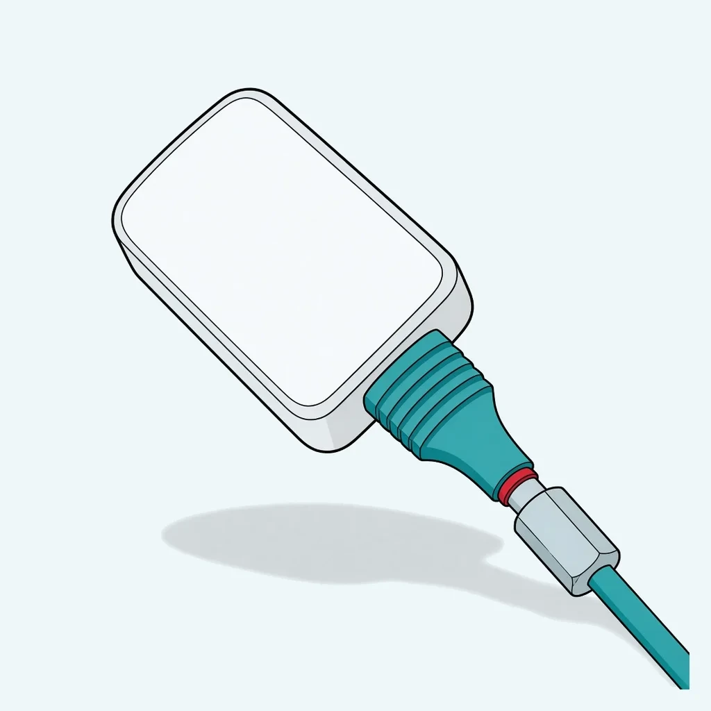
Dental X-ray Sensors: DEXIS, Schick, Carestream, Vatech
X-ray sensor size, cable, and software lock-in, and how DEXIS, Dentsply Sirona, Carestream, and Vatech differ from phosphor plates.
Matching the image to the exam
The FDA’s posting of the 2012 ADA and FDA radiographic selection recommendations treats imaging as an individualized exam, not a standing set of films. For a new adolescent, posterior bitewings are paired with a panoramic examination or with selected periapicals. For a new dentate adult, bitewings are paired with selected periapicals, or with a panoramic examination when it is indicated. Intraoral radiographs are described as more accurate than panoramic radiographs for dentoalveolar trauma, root shape, root resorption, and pulp pathosis. The bitewing examination is called the most efficient method for detecting proximal lesions. A panoramic unit does not retire the sensor or the phosphor plate.
Digital radiography sensors are semicritical in the CDC’s dental sterilization summary. The CDC’s minimum, when a sensor cannot be heat sterilized or high-level disinfected, is an FDA-cleared barrier plus cleaning and an EPA-registered hospital disinfectant with an intermediate-level claim. Follow the sensor maker’s instructions. A camera wand that enters the mouth is in the same semicritical category.
Topics that now land here
X-ray supplies and oral-cancer screening devices now redirect to this page. Owandy’s brand page redirects here as well. A panoramic unit, a sensor, and a CBCT are different machines. The notes below say which image each one makes and which guide compares the hardware.
Sensors and phosphor plates
A digital sensor is the rigid plate on a cable that captures a bitewing or a periapical. A phosphor plate is a flexible plate that is scanned in a reader after the exposure. They make the same kind of intraoral radiograph with different workflow. Sensors are faster to view and cost more to replace when one is dropped. Plates are slower because each plate is read, and a scratched plate is a consumable. The X-ray sensors guide and the phosphor plate guide keep those choices apart. The exposure still comes from an intraoral generator. That machine is the intraoral X-ray units guide.
The CDC treats a digital radiography sensor as semicritical. When the sensor cannot be heat sterilized or high-level disinfected, the minimum is an FDA-cleared barrier plus cleaning and an EPA-registered hospital disinfectant with an intermediate-level claim. Follow the sensor maker’s instructions. Holders have to fit that sensor’s thickness. A holder for a different brand will not seat the same plate. Barriers and positioning kits from the retired X-ray supplies list belong with the sensor or the plate you already own, not as a substitute for either one.
Panoramic units
A panoramic radiograph is the full-arch screening image. The FDA’s posting of the ADA and FDA selection recommendations, already cited above, calls the bitewing the most efficient method for detecting proximal lesions and describes intraoral radiographs as more accurate than panoramic radiographs for dentoalveolar trauma, root shape, root resorption, and pulp pathosis. A panoramic unit does not retire the sensor. It answers a different question: third molars, mixed dentition, and a first look at the jaws. Compare units in the panoramic X-ray guide. A cephalometric attachment is an orthodontic addition. A general office that does not trace cephs does not need the arm.
CBCT
CBCT is a volume, not a sharper bitewing. It is the image for implant planning, impacted teeth, and anatomy a periapical cannot settle. Field of view and voxel size are the spec-sheet items. A larger volume is not a sharper image. A combo unit that also takes a panoramic can replace a separate panoramic machine. It does not replace the sensor for bitewings. Compare systems in the CBCT systems guide.
Intraoral cameras
An intraoral camera records a color picture of a surface the patient can see. It is not a radiograph and it is not an impression scanner. A wand that enters the mouth is semicritical, in the same CDC category as other devices that contact mucous membrane. Compare cameras in the intraoral cameras guide. Do not buy a camera to solve a bitewing problem.
Oral cancer screening devices
Owandy’s retired brand page also lands here. The brand name does not decide whether you need a sensor, a panoramic unit, or a CBCT. Pick the image first, then the machine that makes that image.
Light-based screening devices and similar adjuncts from the retired list are not radiographs. The ADA’s oral cancer guideline says clinicians should take a history and perform an intraoral and extraoral conventional visual and tactile examination in all adult patients. For a suspicious lesion, the guideline says to biopsy or refer. It says salivary and light-based adjuncts are not recommended for evaluating lesions for malignancy. A device does not replace that examination or the decision to biopsy. If you already own one, read what the ADA recommends it not be used for before you treat a color change on the screen as a diagnosis.
What to open first
The retired X-ray supplies list mixed holders, barriers, and plates into one catalog page. Split them when you reorder. A holder positions the receptor. A barrier covers a sensor that cannot be sterilized. A phosphor plate is the receptor. Buying a box of barriers does not give you a sensor, and a scratch on a plate is not fixed by a new holder. Ask the sensor maker which barrier they clear, because a tight barrier on the wrong sensor changes the image and the fit in the holder.
Read the generator and the receptor as a pair. The intraoral unit’s exposure settings have to be ones the sensor or plate instructions allow. A panoramic machine has its own technique factors. A CBCT protocol is chosen by volume, not by the bitewing setting on the wall unit. If the office has one sensor and four rooms, the bottleneck is the sensor, which the sensor guide covers. If the office has a panoramic and still misses proximal lesions, the missing image is the bitewing, which is the point of the FDA and ADA selection statement above.
Everyday bitewings and periapicals start with a sensor or a plate and an intraoral generator. A panoramic is the next machine if you want the full-arch view and you do not yet have a reason for 3D. CBCT comes when the cases need a volume. Cameras and screening lights are communication and adjunct tools, and the ADA statement above is the limit on what a light-based adjunct is for.
Sources
American Dental Association and U.S. Food and Drug Administration
Dental Radiographic Examinations: Recommendations for Patient Selection and Limiting Radiation Exposure. Revised 2012.
Centers for Disease Control and Prevention
Sterilization and Disinfection. Dental Infection Prevention and Control.
Updated May 15, 2024cdc.gov (opens in a new tab)American Dental Association
Oral Cancer Guideline.